Bullous Keratopathy Epidemiology Summary
- Bullous keratopathy is caused by edema of the cornea, resulting from failure of the corneal endothelium to maintain its normally transparent, dehydrated state. Most frequently, it is due to Fuchs corneal endothelial dystrophy or corneal endothelial trauma.
- Angle-closure-related mechanisms emerged as the predominant contributors to bullous keratopathy, accounting for ~51% of cases. In contrast, aphakic and pseudophakic bullous keratopathy were concentrated among cases with missing pre-cataract surgery anterior chamber depth data, highlighting the importance of underlying anterior segment characteristics in bullous keratopathy etiology.
- Corneal endothelial cell density normally ranges between 2,500 and 3,000 cells/mm²; however, corneal decompensation and the development of bullous keratopathy generally occur when the density falls below 500–700 cells/mm².
- Pseudophakic bullous keratopathy is a common indication for corneal transplantation, accounting for approximately 25% of cases across several studies.
- Pseudophakic bullous keratopathy develops in approximately 0.1–2.0% of patients following cataract surgery, although the incidence has declined with modern surgical techniques.
- Japan’s bullous keratopathy etiology profile differs markedly from Western markets, with lens implantation-related cases accounting for a substantially larger share, while Fuchs dystrophy remains relatively uncommon.
- Standardized diagnostic and management guidelines are needed to improve disease identification and treatment consistency, while better understanding of etiology and pathogenesis could support more targeted and potentially curative therapies.
Bullous Keratopathy Epidemiology Forecast in the 7MM
- 2025 Diagnosed Prevalent Cases of Bullous Keratopathy: ~XX
- 2036 Diagnosed Prevalent Cases of Bullous Keratopathy: ~XX
- Bullous Keratopathy Growth Rate (2026–2036): ~XX% CAGR
DelveInsight's ‘Bullous Keratopathy – Epidemiology Forecast – 2036’ report delivers an in-depth understanding of bullous keratopathy, historical and forecasted epidemiology in the United States, EU4 (Germany, Spain, Italy, and France), the United Kingdom, and Japan.
|
Study Period |
2022–2036 |
|
Historical Year |
2022–2025 |
|
Forecast Period |
2026–2036 |
|
Base Year |
2026 |
|
Geographies Covered |
|
|
Bullous Keratopathy Epidemiology CAGR (Study period/Forecast period) |
~XX % (2026–2036) |
|
Bullous Keratopathy Epidemiology Segmentation Analysis |
Patient Burden Assessment
|
Bullous Keratopathy Understanding and Diagnostic Algorithm
Bullous Keratopathy Overview and Diagnosis
Bullous keratopathy is a corneal disorder characterized by corneal edema resulting from irreversible loss or dysfunction of corneal endothelial cells, leading to failure of the endothelial pump mechanism. The condition commonly develops following cataract surgery (pseudophakic bullous keratopathy), ocular trauma, Fuchs endothelial corneal dystrophy, glaucoma surgery, or other intraocular procedures. Progressive corneal edema causes epithelial bullae, blurred vision, pain, photophobia, foreign body sensation, and recurrent epithelial erosions, significantly impairing quality of life. Diagnosis is based on a comprehensive ophthalmic examination using slit-lamp biomicroscopy, supported by specular microscopy to assess endothelial cell density and morphology, corneal pachymetry to measure corneal thickness, and anterior segment optical coherence tomography (AS-OCT) to evaluate the extent of corneal edema. Early and accurate diagnosis is essential to guide appropriate management and determine the need for medical therapy or corneal transplantation.
Further details are provided in the report.
Bullous Keratopathy Epidemiology
Key Findings from Bullous Keratopathy Epidemiological Analysis and Forecast
- Among major etiologies of bullous keratopathy, FECD accounted for the highest number of cases, nearly 70% of the total cases in the US, followed by cataract surgery, which accounted for approximately 25%. On the contrary, those in glaucoma surgery cases were the least, at only 5% of the total cases.
- FECD accounts for the largest share of bullous keratopathy cases (~50%), followed by cataract surgery (~10%) and glaucoma surgery (~40%).
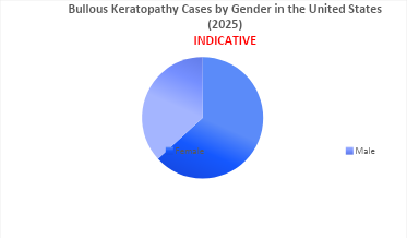
- Bullous keratopathy demonstrates a slight female predominance, with females accounting for approximately 60% of cases compared with 40% in males.
- In the US, ~80% of corneal transplant/keratoplasty cases are overall cases, while ~20% are attributable to bullous keratopathy.
- Bullous keratopathy predominantly affects older adults, reflecting its close association with age-related endothelial cell loss and cataract surgery.
- Approximately 30–40% of patients undergoing endothelial keratoplasty for bullous keratopathy have underlying Fuchs endothelial corneal dystrophy, highlighting it as one of the leading predisposing conditions for endothelial decompensation requiring surgical intervention.
Scope of the Report
- The report covers a segment of an executive summary, a descriptive overview of bullous keratopathy, explaining its causes, signs and symptoms, and pathogenesis.
- Comprehensive insight has been provided into the epidemiology segments and forecasts, the future growth potential of the diagnosis rate, and disease progression.
Report Insights
- Bullous Keratopathy Patient Population Forecast
Report Key Strengths
- Epidemiology‑based (Epi‑based) Bottom‑up Forecasting
- 11-year Forecast
- Patient Burden Trends (By Geography)
FAQs
- What are the disease risks, burdens, and unmet needs of bullous keratopathy? What will be the growth opportunities across the 7MM concerning the patient population with bullous keratopathy?
- What is the historical and forecasted bullous keratopathy patient pool in the US, EU4 (Germany, France, Italy, and Spain), the UK, and Japan?
Reasons to Buy
- Insights on patient burden/disease prevalence, evolution in diagnosis, and factors contributing to the change in the epidemiology of the disease during the forecast years.
- To understand key opinion leaders’ perspectives on the diagnostic challenges to overcome barriers in the future.
- Detailed insights on various factors hampering disease diagnosis and other existing diagnostic challenges.